What Your Nails Say About Your Health
Introduction
Nails are often overlooked in daily medical assessment, yet they are windows into our inner health. Subtle changes in their color, shape, thickness, or surface may reflect not only local conditions but also systemic diseases. From nutritional deficiencies to chronic organ dysfunction, nails silently record what the body endures. A physician who trains the eye to read these signs can often detect illness earlier than routine laboratory investigations.
General Readers
For the layperson, nails are more than a cosmetic feature. Healthy nails are generally smooth, uniformly colored, and firm. Warning signs to look out for include:
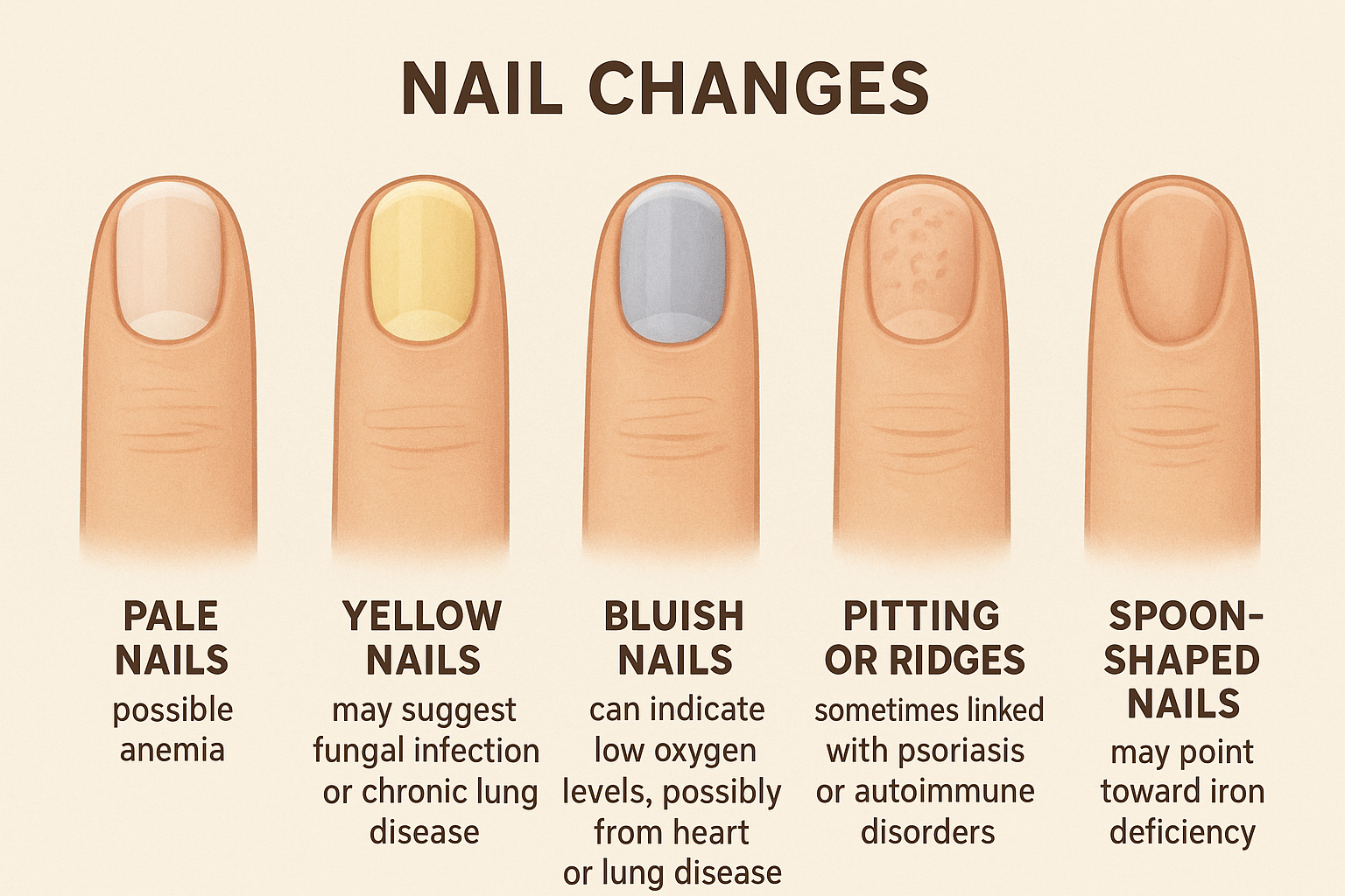
• Pale nails – possible anemia.
• Yellow nails – may suggest fungal infection or chronic lung disease.
• Bluish nails – can indicate low oxygen levels, possibly from heart or lung disease.
• Pitting or ridges – sometimes linked with psoriasis or autoimmune disorders.
• Spoon-shaped nails (koilonychia) – may point toward iron deficiency.
Your nails are a mirror of your internal health. Any persistent change should never be ignored.
Medical Students
For students, nails provide an excellent clinical examination point. They demonstrate the link between systemic pathology and peripheral manifestation. Important associations include:
• Clubbing: bulbous enlargement of the fingertips; classically associated with chronic hypoxemia, lung cancer, interstitial lung disease, cyanotic heart disease, and inflammatory bowel disease.
• Leukonychia: white discoloration, often related to hypoalbuminemia (liver disease, nephrotic syndrome, protein-calorie malnutrition).
• Beau’s lines: transverse depressions across the nail plate, indicating temporary cessation of nail growth after systemic illness, high fever, or chemotherapy.
• Onycholysis: separation of the nail from the nail bed, seen in thyrotoxicosis, psoriasis, or trauma.
• Mee’s lines: transverse white bands, classically linked with arsenic poisoning but also seen in chemotherapy.
Students must learn to integrate these findings into differential diagnosis rather than treating them in isolation.
Young Doctors
For junior clinicians, nails offer a rapid bedside clue during routine patient encounters. An observant doctor can save time and direct investigations wisely. Practical points:
• Always inspect nails in good light as part of general examination.
• Look for symmetry (systemic disease) versus localized changes (trauma, infection).
• In the presence of nail abnormalities, always correlate with systemic findings (pallor, jaundice, clubbing, lymphadenopathy).
• Document nail changes clearly in the case sheet—often they are overlooked in daily rounds.
• Remember: nails grow slowly (about 3 mm/month for fingernails, 1 mm/month for toenails), so nail changes often reflect disease processes of weeks to months’ duration.
General Practitioners
For family physicians and GPs, nails are a part of holistic examination. Patients rarely come with “nail complaints,” but nails can guide you toward underlying diagnoses:
• Repeated respiratory complaints + clubbing → investigate for lung pathology.
• Chronic fatigue + pale, spoon-shaped nails → consider iron studies.
• Yellow nails with lymphedema or pleural effusion → think of Yellow Nail Syndrome.
• Multiple horizontal ridges → inquire about recent major illnesses or stressors.
• Painful, red swelling near the nail (paronychia) → manage as infection but also evaluate for diabetes if recurrent.
For GPs, nail examination is not merely diagnostic—it also builds patient trust, as patients feel the doctor is thorough and attentive.
When to See the Doctor
You should seek medical advice if:
• Your nails change color, shape, or thickness without clear reason.
• Persistent brittleness, splitting, or detachment of nails occurs.
• You notice painful swellings, discharge, or recurrent infections.
• Nails appear clubbed or bluish, suggesting low oxygen.
• Systemic symptoms like fatigue, weight loss, cough, or abdominal swelling accompany nail changes.
Conclusion
Nails, though small and silent, can speak volumes about your health. For the physician, they are a readily visible diagnostic canvas; for the patient, they are a personal health indicator. Observing nails carefully can bring hidden systemic illnesses to light. In modern practice, where machines dominate, the art of bedside clinical observation should never be forgotten. Nails remind us that medicine begins with the eye of the clinician before the lens of the laboratory.
Pathophysiology of Nail Changes
1. Clubbing
• Mechanism:
• Chronic hypoxemia (e.g., from lung or heart disease) leads to vasodilation in the distal digits.
• Increased blood flow and vascular endothelial growth factor (VEGF) promote connective tissue proliferation around the nail bed.
• Hypertrophy of soft tissue occurs, raising the nail plate and causing the characteristic bulbous appearance.
• Key Point: Clubbing reflects chronic tissue hypoxia and abnormal vascular growth factors.
2. Koilonychia (Spoon-shaped nails)
• Mechanism:
• Iron deficiency impairs heme synthesis and reduces oxygen-carrying capacity.
• The resulting hypoxia at the nail matrix weakens keratin formation, leading to nail thinning.
• Over time, nails become concave (spoon-shaped) due to poor structural integrity.
• Key Point: Indicates defective keratinization secondary to iron deficiency anemia.
3. Leukonychia (White nails)
• Mechanism:
• True leukonychia arises from defective keratinization in the nail matrix, trapping air within the nail plate.
• Apparent leukonychia (seen in hypoalbuminemia due to liver disease, nephrotic syndrome, or malnutrition) occurs because the nail bed beneath the plate is pale due to reduced vascularity.
• Key Point: Either matrix abnormality or underlying vascular/nutritional deficiency leads to the white appearance.
4. Beau’s Lines
• Mechanism:
• Severe systemic stress (high fever, chemotherapy, sepsis, trauma) temporarily halts nail matrix proliferation.
• When recovery begins, growth resumes, leaving a transverse groove across the nail plate.
• Key Point: Represents temporary cessation of nail growth due to systemic illness.
5. Onycholysis (Nail plate separation)
• Mechanism:
• Hyperthyroidism → increased keratinocyte turnover and fragile nail bonds.
• Psoriasis → inflammatory cytokines damage nail bed epithelium, loosening attachment.
• Trauma or infection → direct structural damage.
• Key Point: Results from loss of adhesion between nail bed and nail plate.
6. Mee’s Lines (Transverse white bands)
• Mechanism:
• Arsenic poisoning or chemotherapy damages the nail matrix, causing transverse leukonychia.
• As the nail grows, the band moves distally, reflecting the period of toxic insult.
• Key Point: Sign of matrix injury from toxins.
7. Yellow Nails
• Mechanism:
• In fungal infection: dermatophytes produce keratinases, degrading keratin and causing thickened, yellow nails.
• In systemic disease (Yellow Nail Syndrome): impaired lymphatic drainage and protein accumulation discolor the nails.
• Key Point: Caused by keratin degradation or lymphatic dysfunction.
8. Bluish Nails (Cyanosis)
• Mechanism:
• Reduced oxygen saturation of hemoglobin (<85%) leads to bluish discoloration.
• The nail bed, being thin and translucent, clearly reflects venous blood color.
• Key Point: Direct indicator of hypoxemia or poor peripheral circulation.
9. Pitting of Nails
• Mechanism:
• Seen in psoriasis and alopecia areata due to defective keratinization in the proximal nail matrix.
• Keratinocyte clusters fall out, leaving small depressions on the nail surface.
• Key Point: Matrix inflammation and disordered keratin formation create pits.
Summary
Nail changes occur because the nail matrix and nail bed are highly sensitive to systemic oxygenation, nutritional status, toxins, vascular supply, and inflammatory cytokines. Since nails grow slowly, their abnormalities often represent disease processes over weeks to months, making them reliable biological markers of chronic illness.